New Studies
Genetics of Cleft Lip and Palate:
A new study provided by The National Institutes of Health, shows that there are two human genes identified as a factor in causing cleft lip and/or palate, when they are found in a slightly altered form. These genes are called, MAFB and ABCA4. It has also been stated, that people born with clefts seem to have frequent alterations in IRF6 gene and a suspected segment of chromosome eight. These new findings will be an important boost forward in this field.
New Treatment For Cleft Lip & Palate:
Philip Stoddard and Beth Roscoe of Shriners Hospitals for Children, created a new method of pre-surgical treatment for infants born with wide clefts of the lip and palate by narrowing the gap between the gingivas prior to cleft lip surgery.
There are many essential benefits in undergoing this procedure:
A new study provided by The National Institutes of Health, shows that there are two human genes identified as a factor in causing cleft lip and/or palate, when they are found in a slightly altered form. These genes are called, MAFB and ABCA4. It has also been stated, that people born with clefts seem to have frequent alterations in IRF6 gene and a suspected segment of chromosome eight. These new findings will be an important boost forward in this field.
New Treatment For Cleft Lip & Palate:
Philip Stoddard and Beth Roscoe of Shriners Hospitals for Children, created a new method of pre-surgical treatment for infants born with wide clefts of the lip and palate by narrowing the gap between the gingivas prior to cleft lip surgery.
- A plaster model of the patient's palate is made from a dental impression.
- Plaster model is then scanned using a 3-D laser scanner.
- Corrections are designed to gradually reduce the gap, while accommodating growth by using CAD/CAM (computer-assisted design and computer-assisted manufacturing) and a series of palate-shape corrections are digitally modeled.
- A series of corrective appliances are made for the patient. Each appliance is worn for one week, and is then exchanged for the next one in sequential order. Generally, the entire series is completed within 10 to 15 weeks.
- The patient has then completed the pre-surgical treatment, and may start main surgery.
There are many essential benefits in undergoing this procedure:
- The pattern of molding allows for expected growth.
- Appliances are non-invasive.
- Able to reduce the number of visits to the hospital by be giving the family several appliances in the series at a time.
- Time saving compared to prior methods of pre-surgical treatment.
Dental placodes and their role in tooth formation
Biological tooth replacement
Tooth agenesis
- The earliest morphological sign of tooth formation is the appearance of the primary dental laminae, which are stripes of thickened epithelium, marking the future rows of teeth.
- Within the primary dental lamina placodes form which resemble morphologically, as well as in their molecular regulation, the placodes found during the development of other ectodermal organs.
- The placodes consist of thickened epithelium and underlying neural crest derived ectomesenchyme, and they function as the first signaling centers of the tooth.
- One particular hypothesis suggests that each dental placode gives rise to an entire tooth family (incisor, canine, molar).
- The epithelium first buds into the mesenchyme and then grows to encompass the mesenchymal dental papilla during the cap stage.
- The epithelial cervical loop is formed during the cap stage on the lateral sides of the bud. Different developmental choices are made at this point leading to the formation of different tooth types.
- The epithelial stem cell niche in the cervical loop is maintained in continuously growing teeth, but disappears in teeth which develop roots such as all human teeth.
Biological tooth replacement
- Replacement of missing or damaged teeth currently involves fixed or removable prostheses or dental implants.
- Dental implants are increasing within these years.
- Dental implants involve drilling a hole into the jawbone into which titanium rod is screwed that is capped by a plastic or ceramic tooth crown.
- Implants are possible only when there is sufficient bone into which titanium rod can be integrated.
Tooth agenesis
- Death of a tooth.
- Supernumary teeth occur much less frequently than agenesis.
- Occurs most frequently on the maxillary central and lateral incisors, wisdom teeth and second premolar.
- Permanent teeth are more affected than primary teeth.
- If the primary tooth is missing its permanent teeth will also be missing.
- Tooth agenesis originates from either genetic or environmental factors.
- Genetic: familial tooth agenesis is transmitted as an autosomal dominant, autosomal recessive, or X-linked genetic condition. There are more than 49 syndromes associated with tooth agenesis.
- Environmental: virus infection, toxins and radio- or chemotherapy, trauma.
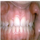
Intra oral photograph of anterior arch form of a 16 year-old female patient with bilateral agenesis of laterral incisers, whose canines were moved mesially into the lateral inciser position. (1)
Reference:
(1) Arvystas Michael: Orthodontic management of genesis and other complexities. London: Martin Dunitz Ltd, member of the Taylor & Francis plc, 2003, page:23.
(1) Arvystas Michael: Orthodontic management of genesis and other complexities. London: Martin Dunitz Ltd, member of the Taylor & Francis plc, 2003, page:23.