General Embryology
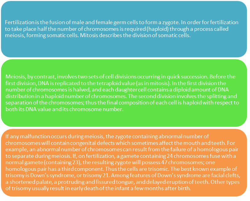
Germ Cell Formation and Fertilization:
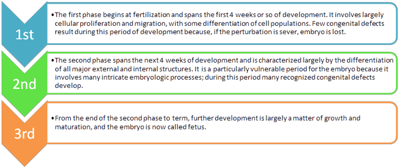
Prenatal Development:
Prenatal development is divided into 3 successive phases:
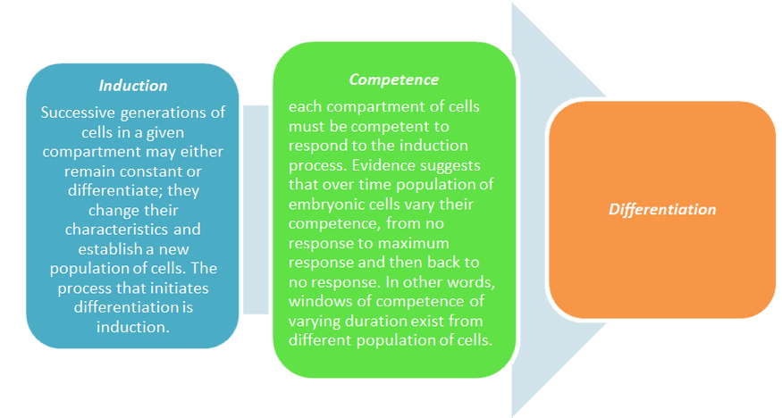
Induction, Competence, and Differentiation:
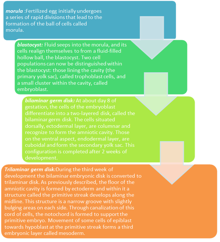
Formation
of the Three-Layered Embryo:
Formation of the neural tube and fate of the germ layers:
- In the first 3 weeks of the embryo development, embryo has been sketch
"proliferation and migration".
- The next 3 to 4 weeks of development differentiation of major tissues
and organs occur "differentiation".
- Nervous system and neural crest tissue differentiate from ectoderm.
- Nervous system develop as thickening within the ectoderm layer "
Neural plate "
- Rapidly form raised margins "Neural Folds ".
- Neural folds encompass forming "Neural groove".
- Neural folds fuse and form "Neural tube" that form the floor
of amniotic cavity.
- Thickening of mesoderm to form paraxial mesoderm that break into blokes
to form "somite" which is compose of
"sclerotome,myotome,dermatome", periphery of the paraxial mesoderm
there is intermediate mesoderm ,which become the urogenital system.
- Thickening of the lateral mesoderm give rise to connective tissue
associated with muscle and viscera.
- In the head the neural tube undergoes massive expansion to form some
parts of the brain.
Folding of the embryo:
- Along the rostrocaudal axis, the head folds to form primitive stomodeum which
line by ectoderm, separated the gut by "buccopharyngeal membrane"
- Along the lateral axis, the ectoderm of the aminiotic cavity encapsulates the embryo and forms the surface epithelium
The Neural crest:
- Differentiated and separated from the crest of the neural folds
- At the neural crest cell's induction phase, cells undergo epithelial mesenchymal transformation
- The neural crest cells in the head have an important role by forming cranial sensory ganglia and most of the connective tissue of the head
- All the tissues of the tooth (except
enamel and some cementum) develop from the neural crest cells that provide proper
dental development